Lesson 4 of 7 / Cells and stem cells
Read a micrograph without guessing
Which visible evidence supports the label?
In this lesson: Use real light and electron micrographs alongside drawings to identify structures and state limits.
About 5 min
The key ideaUse several visible features and the image scale. A tidy textbook colour is not an identification test.
Try each image before revealing its explanation. The light images show whole-cell organisation; the electron images resolve finer membrane patterns.
Explore the idea
Start with the visible evidence
Look at real cells
Find a cell wall and a nucleus. Can you distinguish the membrane separately from the wall?
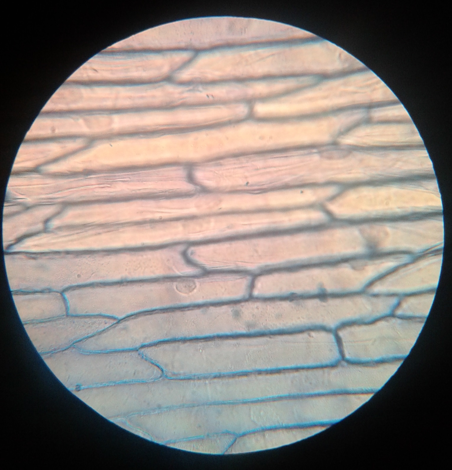
Image size changes with your screen. Use a stated scale bar for measurements; do not calculate from the display size alone. Practise slide preparation and focusing in your laboratory.
Explanation
A light micrograph often shows a cell outline, cytoplasm, a stained nucleus and, in suitable plant tissue, chloroplasts. Many thin membranes and small ribosomes cannot be resolved individually. A boundary visible in a plant image may be a wall rather than a separately resolved membrane.
Electron micrographs reveal finer structures but usually show a thin section through a three-dimensional object. An organelle can therefore appear circular, elongated or incomplete depending on the cutting plane. Staining produces contrast, not the natural colour of the organelle.
Build an identification from a pattern: rough ER has membranes with attached ribosome dots; a Golgi body has flattened stacks with nearby vesicles; mitochondria have internal cristae. Cytosol occupies spaces around organelles, rather than having its own surrounding membrane.
Distinguish observation from inference. State what is visible, explain what it supports, and acknowledge unresolved details. These images practise interpretation; they do not show that you can prepare specimens or operate a microscope.
Step by step
- 1
Observe
Describe the boundary, internal pattern and neighbouring structures.
- 2
Infer
Match a combination of features to an organelle.
- 3
Qualify
Do not claim an invisible membrane or an exact size without suitable evidence.
Worked example
A dot is not enough
A student identifies every dark dot in an electron micrograph as a ribosome. Improve the method.
One way to explain it
Check the scale and distribution. Numerous very small dots attached along ER membranes support ribosome identification; a single larger dark vesicle could be something else. Use the membrane pattern and surrounding structures as supporting evidence.
Why this answer works
- Recognise the weakness of a single feature.
- Add size, arrangement and context.
Is this true? "Every organelle must look like the outline in the diagram."
A diagram is a model. Section angle, specimen preparation and resolution alter the appearance in a real image.