Lesson 2 of 7 / Cells and stem cells
Find the compartments in a cell
How does an internal boundary help a cell work?
In this lesson: Recognise major eukaryotic structures and explain their functions.
About 6 min
The key ideaCell structures create useful spaces and surfaces. Identify their visible features, then connect those features to a specific function.
Explore the idea
Match a visible feature to a job
Visible clue: A large compartment with a double envelope; the darker nucleolus is a region inside it. Function: DNA is housed in the nucleus. The nucleolus produces rRNA and assembles ribosomal subunits.
Read the real micrograph
Use the existing labels to find the nuclear boundary and the darker nucleolus. Which of these two structures has its own enclosing membrane?
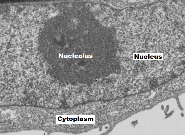
No scale bar is shown in this crop, so it does not support an exact size calculation. Display size changes with your screen; it is not a fixed microscope magnification.
Original schematic. Structures, cell numbers and distances are illustrative, not to scale.
Explanation
The cell surface membrane separates the cytosol from the surroundings and controls exchange. Cytosol is the fluid part of the cytoplasm, where many metabolic reactions occur; cytoplasm commonly includes cytosol and organelles outside the nucleus. A plant cellulose wall lies outside the membrane and provides mechanical support.
The nucleus contains most of a eukaryotic cell's DNA. Its double nuclear envelope separates nuclear contents from cytoplasm; pores allow regulated exchange. The darker nucleolus produces ribosomal RNA and assembles ribosomal subunits. The nucleus and nucleolus are not interchangeable labels.
Mitochondria have an outer membrane and an inner membrane folded into cristae. They support aerobic respiration and ATP production. Chloroplasts contain internal thylakoid membranes, often stacked as grana, and carry out photosynthesis. Both are bounded organelles, unlike a ribosome.
Centrioles are cylinders made from microtubules, commonly present as a pair in an animal-cell centrosome. They contribute to organising the spindle during division. A section across a cylinder looks different from a section along it, so do not rely on one outline alone when interpreting a micrograph.
Step by step
- 1
Look for boundaries
Decide whether the feature is outside the cell, inside it, or surrounding the nucleus.
- 2
Look inside the boundary
Cristae, membrane stacks or a darker nucleolus help distinguish structures.
- 3
Give a specific job
For example, link a mitochondrion to aerobic respiration, not simply to being important.
Worked example
Two internal membrane patterns
An image shows an oval with inner membrane folds and a second oval with stacks of internal membranes. Suggest their identities in a leaf cell.
One way to explain it
The first is consistent with a mitochondrion because of its cristae. The second is consistent with a chloroplast because of its stacked thylakoids. The tissue context supports, but does not replace, those visible clues.
Why this answer works
- Use internal structure, not colour in a stained image.
- Give the feature that supports each identification.
Is this true? "The nucleolus is a small nucleus with its own membrane."
The nucleolus is a region within the nucleus and is not membrane-bound. It is associated with ribosomal RNA production and subunit assembly.