Lesson 1 of 9 / Nutrition and transport in flowering plants
Leaf structure and gas exchange
How does a leaf bring carbon dioxide close to its chloroplasts?
In this lesson: Identify tissues in a dicot leaf section and explain their roles in photosynthesis and gas exchange.
About 6 min
The key ideaStomata and mesophyll air spaces shorten the diffusion route to photosynthesising cells; chloroplast-rich palisade tissue captures light.
Palisade mesophyll: Closely packed elongated cells with many chloroplasts, near the illuminated upper surface.
Brown ring: selected feature. Dark green ovals: chloroplasts. Blue/gold in the vein: xylem/phloem. Original schematic of a typical dicot section, not a microscope photograph; sizes and cell numbers are simplified.
Read a real microscope section
Find the upper epidermis, palisade tissue and spongy tissue. Which visible pattern suggests a route for gases within the leaf?
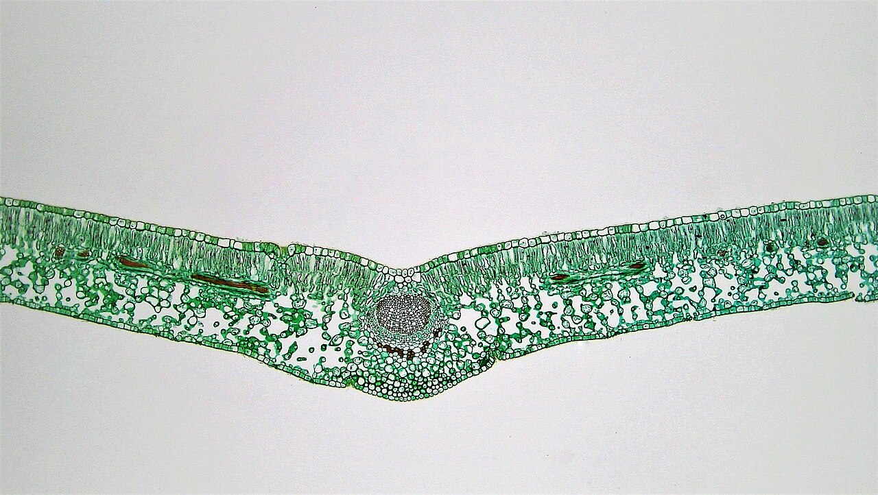
Image size changes with your screen. Use a stated scale bar for measurements; do not calculate from display size alone. Practise specimen observation and focusing in your laboratory.
Explanation
A typical dicot leaf has an upper epidermis, palisade mesophyll, spongy mesophyll and lower epidermis. A transparent epidermis lets light reach the mesophyll. A waxy cuticle reduces water loss. Palisade cells lie near the illuminated upper surface and contain many chloroplasts.
Spongy mesophyll has irregularly arranged cells with large air spaces. These allow gases to spread through the leaf and provide a large moist cell surface for gas exchange. Spongy cells also contain chloroplasts, though usually fewer than palisade cells. Most epidermal cells lack chloroplasts; guard cells are an exception.
Stomata are pores controlled by pairs of guard cells, often concentrated on the lower epidermis in a typical terrestrial dicot leaf. During net photosynthesis, carbon dioxide diffuses through open stomata into air spaces, dissolves in the moist surfaces of mesophyll cell walls and diffuses into the cells, reaching chloroplasts where it is used.
A vascular bundle contains xylem supplying water and mineral ions and phloem transporting food. In a leaf section, connect each structure to a process: chloroplast distribution to photosynthesis, stomata and air spaces to gas exchange, and vascular tissue to transport. Compare the original schematic with the real privet leaf micrograph. Use the arrangement of cells and air spaces as evidence; actual sections vary in shape and detail.
Step by step
- 1
Orient the section
Find the upper epidermis and the palisade layer below it.
- 2
Trace the gas route
Follow carbon dioxide from a stoma through air spaces into mesophyll cells.
- 3
Link structure to function
Explain how the arrangement supports light absorption, diffusion or transport.
Worked example
An irregular tissue with air spaces
A light-microscope section shows irregular cells separated by large spaces below the palisade layer. Identify the tissue and explain a function.
One way to explain it
It is spongy mesophyll. Its interconnected air spaces allow gases such as carbon dioxide to diffuse through the leaf towards mesophyll cells, where carbon dioxide is used in photosynthesis.
Why this answer works
- Use position and arrangement together.
- Name the tissue rather than just "air holes".
- Link the spaces to diffusion and photosynthesis.
Is this true? "Carbon dioxide enters the xylem and is carried to each chloroplast."
Carbon dioxide reaches mesophyll cells mainly by diffusion through stomata and air spaces. Xylem transports water and mineral ions.