Lesson 2 of 5 / Cells and microscopy
Follow a protein through a cell
Why does a protein-secreting cell have extensive rough ER and Golgi?
In this lesson: Recognise and connect ribosomes, ER, Golgi, lysosomes and membranes.
About 7 min
The key ideaRibosomes make polypeptides; the ER and Golgi process and route suitable products in membrane vesicles.
Explore the idea
Follow a protein through compartments
Rough ER: Ribosomes on the cytosolic surface make polypeptides that enter the ER. The ER lumen provides an environment for folding and early processing.
This tracks a selected protein cargo, not movement of whole organelles. Smooth ER lacks attached ribosomes and contributes to lipid synthesis and detoxification. Free ribosomes also synthesise proteins, including many used in the cytosol.
Try a real electron micrograph
Use the photograph on the right. Which internal pattern matches the diagram?
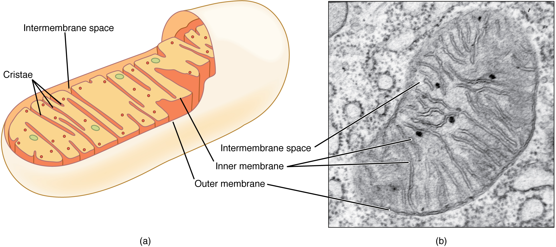
Different section angles change how an organelle looks. Match several visible features before naming it.
Identify a real lysosome
Read the real micrograph
Find the large, irregular dark compartment near the centre. Compare its varied contents with the internal folds of nearby mitochondria.
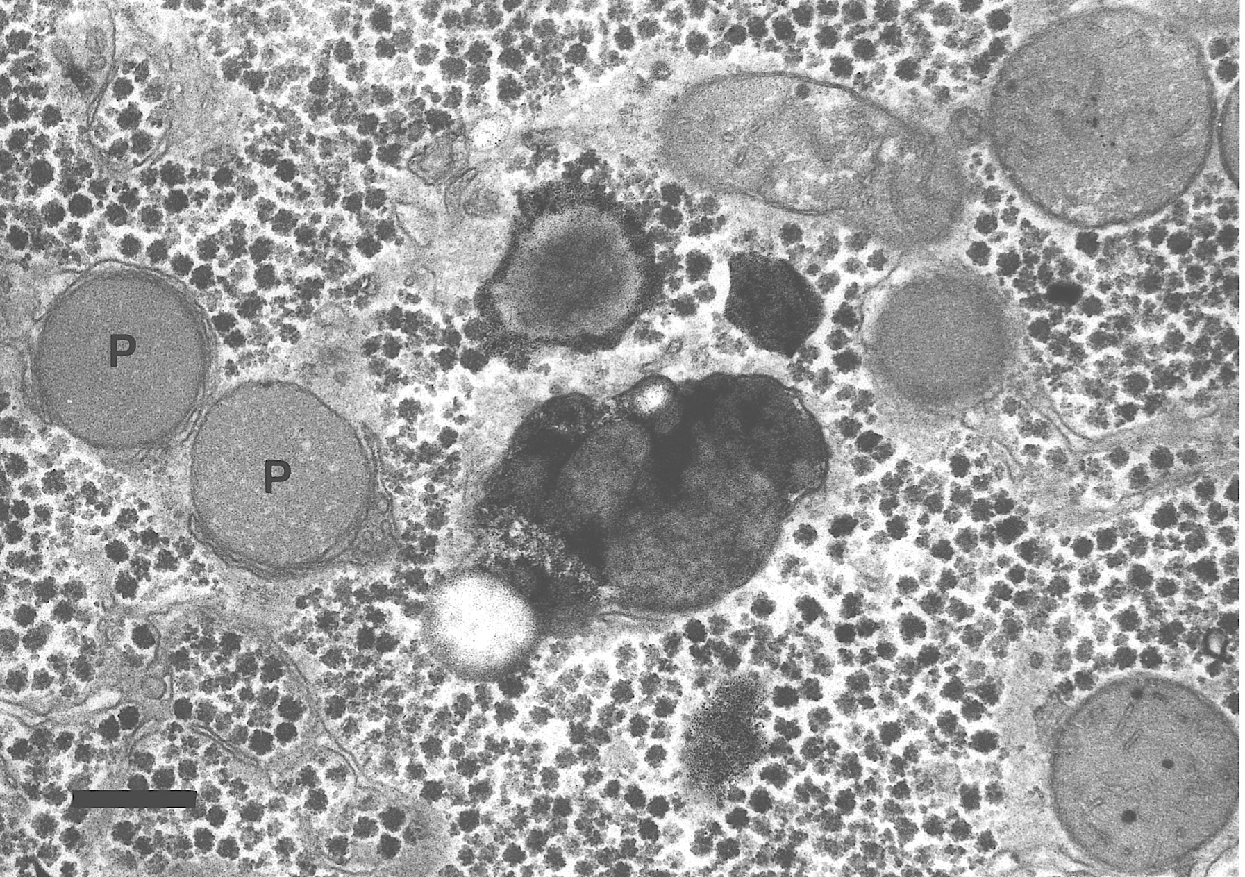
The source identifies the black bar as 0.5 micrometres. Peroxisomes provide context here; their detailed roles are outside this lesson. Display size changes with your screen; it is not a fixed microscope magnification.
Original schematic. Features, cell sizes and distances are not to scale. Use several visible clues when interpreting an unfamiliar section.
Explanation
The cytosol is the aqueous component of the cytoplasm in which many reactions occur. Ribosomes are non-membrane-bound assemblies of rRNA and protein that translate mRNA. Free ribosomes generally make proteins used in the cytosol; ribosomes attached to rough endoplasmic reticulum make many secreted and membrane proteins.
Rough ER consists of membrane cisternae with ribosomes on the cytosolic surface. A growing suitable polypeptide enters the ER, where folding and initial processing occur. Vesicles carry it to the Golgi body, a stack of flattened sacs. The Golgi modifies, sorts and packages products for particular destinations.
A secretory vesicle fuses with the cell surface membrane and releases its contents by exocytosis. Lysosomes are membrane-bound compartments containing hydrolytic enzymes. Their membrane separates digestion of worn components or ingested material from the cytosol; they are not protein-making organelles.
Smooth ER is a membrane network without attached ribosomes and has roles in lipid synthesis and detoxification. In a micrograph, compare several clues: dots on cisternae suggest rough ER; a curved stack with nearby vesicles suggests Golgi. Section angle and staining can change appearances, so a single dark dot is insufficient evidence.
Step by step
- 1
Locate synthesis
Find ribosomes on the cytosolic ER surface.
- 2
Follow membrane traffic
Trace ER to vesicle to Golgi to destination.
- 3
Support an identification
Use membrane organisation and nearby structures, not colour alone.
Worked example
Work through the evidence
A labelled amino acid appears first in rough ER, then Golgi, then extracellular protein. What does this sequence support?
One way to explain it
It supports synthesis and processing through the secretory pathway: ribosomes at rough ER, vesicular transfer to Golgi, then exocytosis.
Why this answer works
- The label tracks newly made protein, not an organelle moving bodily across the cell.
- Timing supports a route; appropriate controls are still needed to exclude alternative interpretations.
Is this true? "The Golgi makes the polypeptide chain."
Ribosomes synthesise the chain. Golgi enzymes modify and sort products already made.