Lesson 3 of 5 / Cells and microscopy
Match organelles to their jobs
Which visible features justify an organelle identification?
In this lesson: Identify nuclear, energy-converting and structural cell components.
About 7 min
The key ideaIdentify a structure using its boundary and internal pattern, then connect those features to its function.
Explore the idea
Use the structure as evidence
Look for: A nuclear envelope surrounds the nucleus; pores interrupt the envelope. A dense nucleolus lies inside. Connect to function: The nucleus houses most eukaryotic DNA. The nucleolus makes rRNA and assembles ribosomal subunits; pores regulate exchange with the cytoplasm.
Read the real micrograph
Use the existing labels to find the nuclear boundary and the darker nucleolus. Which of these two structures has its own enclosing membrane?
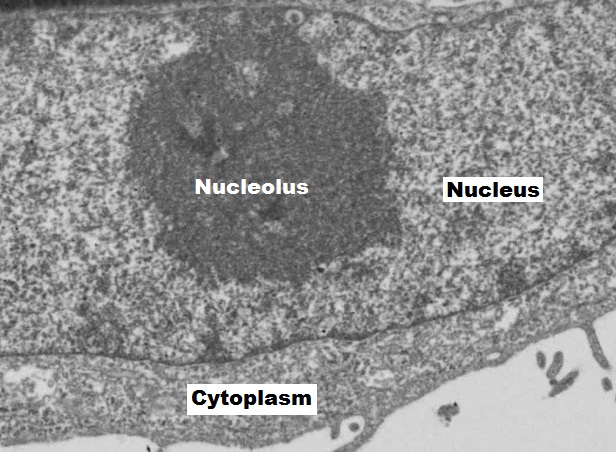
No scale bar is shown in this crop, so it does not support an exact size calculation. Display size changes with your screen; it is not a fixed microscope magnification.
Original schematic. Features, cell sizes and distances are not to scale. Use several visible clues when interpreting an unfamiliar section.
Explanation
The nucleus contains the chromosomes. Its double nuclear envelope has pores through which molecules such as RNA and proteins are exchanged with the cytoplasm. The nucleolus is a dense, non-membrane-bound region where rRNA production and ribosomal subunit assembly occur.
Mitochondria have an outer membrane and an inner membrane folded into cristae. The matrix contains enzymes for stages of aerobic respiration; the inner membrane houses electron carriers and ATP synthase. Cristae provide extensive membrane area for oxidative phosphorylation.
Chloroplasts have an envelope surrounding stroma and internal thylakoid membranes, often stacked as grana. Light-dependent reactions take place at thylakoids, while carbon fixation occurs in the stroma. A plant cell can contain both mitochondria and chloroplasts: photosynthesis does not remove its need for respiration.
The cell surface membrane regulates exchange and communication. A cellulose wall outside a plant cell membrane provides mechanical support and resists excessive expansion. Centrioles are cylindrical microtubule arrangements in many animal cells; within the centrosome they are associated with microtubule organisation during division. Do not add centrioles to every plant cell drawing.
Electron micrographs resolve fine structure that a school light microscope cannot. A nucleus may be visible by light microscopy, but a ribosome, membrane bilayer or the internal architecture of a centriole needs electron microscopy. An identification must match the resolution of the evidence.
Step by step
- 1
Read the boundary
Count membranes where the resolution permits.
- 2
Inspect the interior
Compare cristae, thylakoid stacks or a dense nucleolus.
- 3
Link structure and process
Name the relevant surface or compartment, not only the organelle.
Worked example
Work through the evidence
An organelle has a double boundary and stacks of internal discs. Is it a mitochondrion or chloroplast?
One way to explain it
A chloroplast: the stacks are grana of thylakoids. A mitochondrion instead has inner-membrane folds called cristae.
Why this answer works
- Both can have a double envelope, so that clue alone is insufficient.
- The internal membrane arrangement distinguishes them.
Is this true? "A chloroplast is a plant cell's replacement for a mitochondrion."
Photosynthetic plant cells normally have both. They convert energy through different pathways.