Lesson 5 of 6 / Cell structure and organisation
Organelles seen in finer detail
How does a cell make, package and power its products?
In this lesson: Recognise ribosomes, endoplasmic reticulum, Golgi bodies and mitochondria and state their functions.
About 6 min
The key ideaRibosomes make proteins; the endoplasmic reticulum and Golgi body help process and transport them. Mitochondria release energy through aerobic respiration.
An electron microscope reveals much smaller details than a light microscope. These enlarged drawings show recognition clues; real electron micrographs have varying shapes and orientations.
Explore the idea
Read the pattern, then name the job
Recognition clue: An enclosing boundary with a folded inner membrane.
Function: Aerobic respiration releases energy for cellular activities.
Original diagrams of recognition patterns, not electron micrographs. Structures are shown at different illustrative scales. A real section can cut the same organelle at different angles.
Try a real electron micrograph
Use the photograph on the right. Which internal pattern matches the diagram?
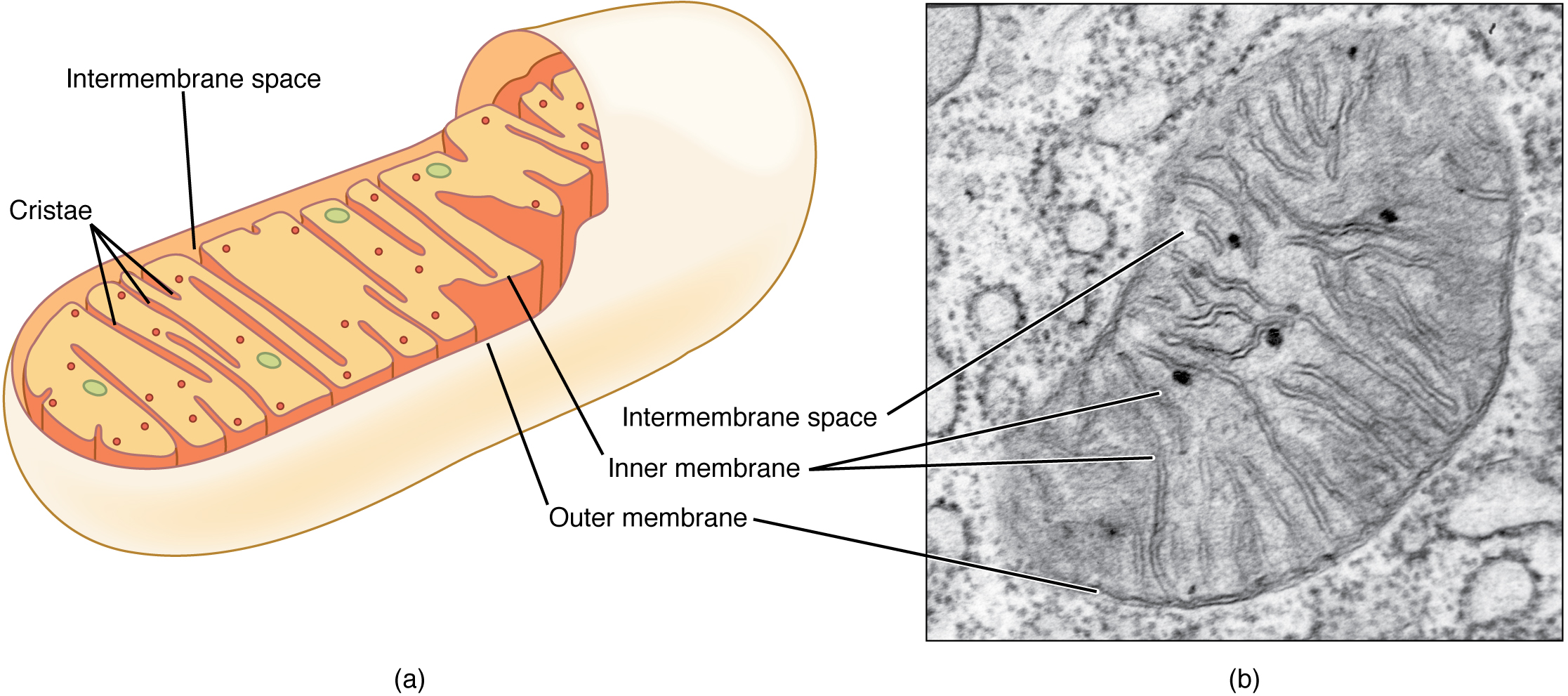
Different section angles change how an organelle looks. Match several visible features before naming it.
Explanation
Ribosomes are small structures where proteins are synthesised from amino acids. They may be free in the cytoplasm or attached to rough endoplasmic reticulum. In an electron micrograph they appear as small dark dots, sometimes clustered or arranged on membranes.
The endoplasmic reticulum is a membrane network within the cell. Rough endoplasmic reticulum has attached ribosomes and is involved in producing and transporting proteins, including proteins to be secreted. Smooth endoplasmic reticulum lacks ribosomes and is involved in making lipids. Look for extended membranes and whether dots are attached.
The Golgi body consists of a stack of flattened membrane sacs, often with small vesicles nearby. It modifies, sorts and packages cell products for transport. A protein that will leave the cell can pass from rough endoplasmic reticulum to the Golgi body, then travel in a vesicle to the membrane.
Mitochondria are sites of aerobic respiration, which releases energy from food substances for cellular activities. The inner membrane is folded. In section, a mitochondrion often appears oval with internal folds, but its apparent shape depends on how it is cut.
Identify structures using several clues. A stack of smooth sacs with surrounding vesicles suggests a Golgi body; a membrane network covered in dots suggests rough endoplasmic reticulum. A dark oval alone is insufficient: check for an enclosing boundary and internal folds before calling it a mitochondrion.
Step by step
- 1
Look for the pattern
Distinguish small dots, extended membranes, stacked sacs and an organelle with internal folds.
- 2
Connect the pattern to the name
Dots on membranes suggest rough endoplasmic reticulum; dots themselves are ribosomes.
- 3
Explain the role
Separate making a protein from modifying and packaging it, and from releasing energy for these activities.
Worked example
A cell that secretes enzymes
A pancreas cell produces and releases protein enzymes. Explain why it has much rough endoplasmic reticulum and a well-developed Golgi body.
One way to explain it
Ribosomes on rough endoplasmic reticulum synthesise the enzyme proteins, which are transported through the endoplasmic reticulum. The Golgi body modifies and packages the proteins into vesicles for secretion.
Why this answer works
- Recognise that these enzymes are proteins.
- Assign synthesis to ribosomes on rough endoplasmic reticulum.
- Assign modification and packaging to the Golgi body.
Is this true? "The Golgi body makes proteins from amino acids."
Ribosomes synthesise proteins. The Golgi body processes and packages proteins after their synthesis.