Lesson 5 of 6 / Cell structure and organisation
Mitochondria and ribosomes
Which organelle releases energy, and which makes protein?
In this lesson: Identify mitochondria and ribosomes in diagrams and electron micrographs and state their functions.
About 5 min
The key ideaMitochondria are sites of aerobic respiration; ribosomes synthesise proteins. A cell needs both processes to function.
Electron micrographs show finer detail than light micrographs. These images are sections through cells, so the same structure can look different at different angles.
Explore the idea
Energy release or protein synthesis?
Use the micrograph below. Find the enclosing boundary and the internal folds.
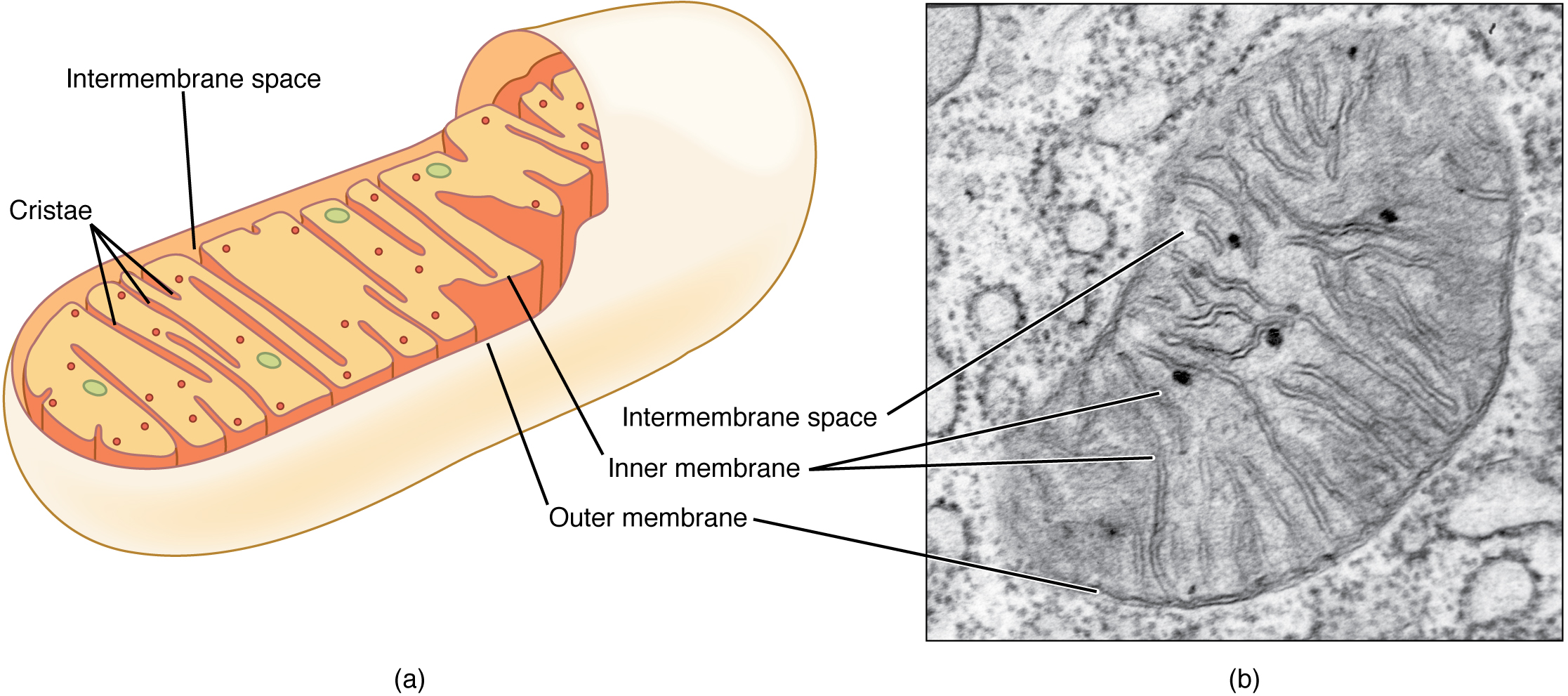
The original diagrams and micrographs have different illustrative scales. A diagram is a model, while a micrograph records a specimen.
Explanation
Mitochondria are organelles in which aerobic respiration occurs. This releases energy from food molecules for cell activities. Cells that have high energy demands, such as contracting muscle cells, commonly contain many mitochondria.
In an electron micrograph, look for an enclosing boundary with an inner membrane folded inside it. The folds are useful evidence for a mitochondrion. An oval outline alone is not enough: different organelles can have similar overall shapes.
Ribosomes are much smaller structures that synthesise proteins from amino acids. In electron micrographs they appear as small dense dots, either in the cytoplasm or on membranes. They are not normally resolved individually using a school light microscope.
A cell making protein needs ribosomes for synthesis and energy released in respiration for its activities. Do not swap these functions: a mitochondrion does not manufacture all the proteins a cell needs, and a ribosome is not the site of aerobic respiration.
Step by step
- 1
Inspect the image
Look for internal folds or a repeated pattern of small dense dots.
- 2
Name the structure
Distinguish the mitochondrion from the much smaller ribosomes.
- 3
State its job
Connect respiration to energy release and ribosomes to protein synthesis.
Worked example
An active muscle cell
A muscle cell has many organelles with folded inner membranes. Identify them and explain why they are useful.
One way to explain it
They are mitochondria. They provide a large capacity for aerobic respiration, releasing energy for repeated muscle contraction.
Why this answer works
- Use the internal folds as identification evidence.
- Connect the named organelle to respiration.
- Link energy release to the specific activity: contraction.
Is this true? "Ribosomes release energy while mitochondria make proteins."
Ribosomes synthesise proteins. Mitochondria are sites of aerobic respiration, releasing energy for cell activities.