Lesson 3 of 5 / Cell structure and organisation
Reading a light microscope view
Why does a real cell look less tidy than a textbook diagram?
In this lesson: Use visible evidence to identify structures and explain how slide preparation improves a view.
About 5 min
The key ideaA stain improves contrast; it does not add a new organelle. Identify what the image actually resolves and distinguish an observation from an inference.
A micrograph is a photograph made through a microscope. Compare real stained onion cells, cheek cells and photosynthetic leaf cells below. First describe what you can see, then check the explanation.
Explore the idea
What can you actually see?
Look at real cells
Find a cell wall and a nucleus. Can you distinguish the membrane separately from the wall?
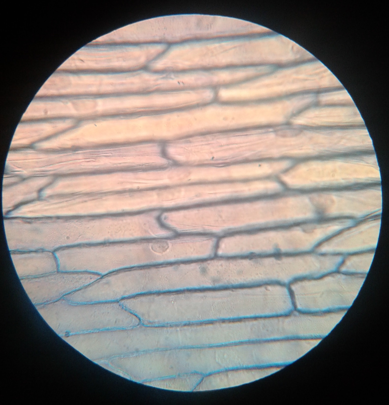
Image size changes with your screen. Use a stated scale bar for measurements; do not calculate from the display size alone. Practise slide preparation and focusing in your laboratory.
Explanation
A biological drawing separates structures clearly. A light microscope view contains overlapping cells, uneven staining and structures at different depths. Start by finding repeated cell boundaries, then look for a nucleus and the surrounding cytoplasm. Only label a structure when the evidence supports it.
For a temporary onion epidermis preparation, a thin layer of epidermis is placed flat in a drop of water on a slide. A suitable stain, such as iodine solution under the instructed school procedure, increases contrast. Lowering a coverslip at an angle helps reduce trapped air bubbles. Handle glass and stains using the laboratory instructions.
Use low power first to locate and centre the specimen. Adjust illumination and focus, then increase magnification if needed. At higher power use fine focus carefully. Higher magnification makes the image larger but cannot reveal detail beyond the resolving power of the microscope.
In onion bulb epidermis, walls form a repeated pattern. A stained nucleus may lie near the edge because a large vacuole occupies much of the cell. The membrane can be hard to distinguish separately from the wall when pressed closely against it. Onion bulb cells normally lack chloroplasts; their absence is expected.
With an appropriate prepared animal slide, look for a membrane boundary, cytoplasm and stained nucleus without a cellulose wall. Light microscope observations should be compared with diagrams, but the diagrams must not replace the evidence. Ribosomes and internal membrane detail require electron microscopy.
Step by step
- 1
Prepare for a clear view
Use a thin, flat specimen, an appropriate stain and a carefully lowered coverslip.
- 2
Observe before naming
Record visible boundaries and darker bodies. A large round air bubble is not automatically a nucleus.
- 3
Recognise the limit
Do not claim that a structure is absent simply because it cannot be resolved or stained clearly.
Worked example
Before and after staining
Before staining, an onion cell has a clear outline but no obvious nucleus. After staining, a dark body becomes visible near its edge. Explain the change.
One way to explain it
The stain increased contrast between the nucleus and its surroundings, making an existing structure easier to see. It did not create a nucleus. The nucleus can be near the edge because the large vacuole occupies much of the cell.
Why this answer works
- Separate visibility from presence.
- Explain the purpose of staining.
- Use plant cell structure to interpret the position.
Is this true? "If I cannot see a cell membrane, the cell has no membrane."
A cell membrane may be too thin or too close to the wall to distinguish separately in this view. Limited resolution or contrast is not proof of absence.